Greater Trochanter on:
[Wikipedia]
[Google]
[Amazon]
 The greater trochanter of the
File:Gray244.png, Right femur. Anterior surface.
File:Gray245.png, Right femur. Posterior surface.
File:Gray339.png, Right hip-joint from the front.
File:Gray343.png, Capsule of hip-joint (distended). Posterior aspect.
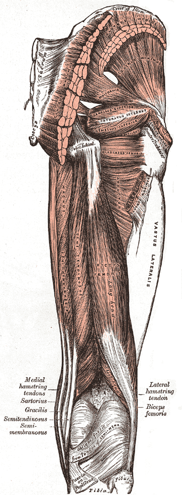
File:Gray544.png, The arteries of the gluteal and posterior femoral regions.
File:Slide2DAD.JPG, Hip joint. Lateral view.Greater trochanter.
 The greater trochanter of the
 femur
The femur (; ), or thigh bone, is the proximal bone of the hindlimb in tetrapod vertebrates. The head of the femur articulates with the acetabulum in the pelvic bone forming the hip joint, while the distal part of the femur articulates with ...
is a large, irregular, quadrilateral eminence and a part of the skeletal system.
It is directed lateral and medially and slightly posterior. In the adult it is about 2–4 cm lower than the femoral head
The femoral head (femur head or head of the femur) is the highest part of the thigh bone (femur). It is supported by the femoral neck.
Structure
The head is globular and forms rather more than a hemisphere, is directed upward, medialward, and a l ...
.Standring, Susan, editor. ''Gray’s Anatomy: The Anatomical Basis of Clinical Practice''. Forty-First edition, Elsevier Limited, 2016, p. 1327. Because the pelvic outlet in the female is larger than in the male, there is a greater distance between the greater trochanter
A trochanter is a tubercle of the femur near its joint with the hip bone. In humans and most mammals, the trochanters serve as important muscle attachment sites. Humans are known to have three trochanters, though the anatomic "normal" includes ...
s in the female.
It has two surfaces and four borders. It is a traction epiphysis
The epiphysis () is the rounded end of a long bone, at its joint with adjacent bone(s). Between the epiphysis and diaphysis (the long midsection of the long bone) lies the metaphysis, including the epiphyseal plate (growth plate). At the jo ...
.
Surfaces
The ''lateral surface'', quadrilateral in form, is broad, rough, convex, and marked by a diagonal impression, which extends from the postero-superior to the antero-inferior angle, and serves for the insertion of the tendon of thegluteus medius
The gluteus medius, one of the three gluteal muscles, is a broad, thick, radiating muscle. It is situated on the outer surface of the pelvis.
Its posterior third is covered by the gluteus maximus, its anterior two-thirds by the gluteal aponeu ...
.
Above the impression is a triangular surface, sometimes rough for part of the tendon of the same muscle, sometimes smooth for the interposition of a bursa between the tendon and the bone. Below and behind the diagonal impression is a smooth triangular surface, over which the tendon of the gluteus maximus
The gluteus maximus is the main extensor muscle of the hip. It is the largest and outermost of the three gluteal muscles and makes up a large part of the shape and appearance of each side of the hips. It is the single largest muscle in the human ...
lies, a bursa
( grc-gre, Προῦσα, Proûsa, Latin: Prusa, ota, بورسه, Arabic:بورصة) is a city in northwestern Turkey and the administrative center of Bursa Province. The fourth-most populous city in Turkey and second-most populous in the ...
being interposed.
The ''medial surface'', of much less extent than the lateral, presents at its base a deep depression, the trochanteric fossa
In mammals including humans, the medial surface of the greater trochanter has at its base a deep depression bounded posteriorly by the intertrochanteric crest, called the trochanteric fossa. This fossa is the point of insertion of four muscles. Mo ...
(digital fossa), for the insertion of the tendon of the obturator externus
The external obturator muscle, obturator externus muscle (; OE) is a flat, triangular muscle, which covers the outer surface of the anterior wall of the pelvis.
It is sometimes considered part of the medial compartment of thigh, and sometimes c ...
, and above and in front of this an impression for the insertion of the obturator internus
The internal obturator muscle or obturator internus muscle originates on the medial surface of the obturator membrane, the ischium near the membrane, and the rim of the pubis.
It exits the pelvic cavity through the lesser sciatic foramen.
The i ...
and superior and inferior gemellus muscle
The gemelli muscles are the inferior gemellus muscle
and the superior gemellus muscle, two small accessory fasciculi to the tendon of the internal obturator muscle. The gemelli muscles belong to the lateral rotator group of six muscles of the h ...
s.
Borders
The ''superior border'' is free; it is thick and irregular, and marked near the center by an impression for the insertion of thepiriformis
The piriformis muscle () is a flat, pyramidally-shaped muscle in the gluteal region of the lower limbs. It is one of the six muscles in the lateral rotator group.
The piriformis muscle has its origin upon the front surface of the sacrum, and in ...
.
The ''inferior border'' corresponds to the line of junction of the base of the trochanter with the lateral surface of the body; it is marked by a rough, prominent, slightly curved ridge, which gives origin to the upper part of the vastus lateralis
The vastus lateralis (), also called the vastus externus, is the largest and most powerful part of the quadriceps femoris, a muscle in the thigh. Together with other muscles of the quadriceps group, it serves to extend the knee joint, moving the ...
.
The ''anterior border'' is prominent and somewhat irregular; it affords insertion at its lateral part to the gluteus minimus
The gluteus minimus, or glutæus minimus, the smallest of the three gluteal muscles, is situated immediately beneath the gluteus medius.
Structure
It is fan-shaped, arising from the outer surface of the ilium, between the anterior and infer ...
.
The ''posterior border'' is very prominent and appears as a free, rounded edge, which bounds the back part of the trochanteric fossa
In mammals including humans, the medial surface of the greater trochanter has at its base a deep depression bounded posteriorly by the intertrochanteric crest, called the trochanteric fossa. This fossa is the point of insertion of four muscles. Mo ...
.
Additional images
See also
*Third trochanter
In human anatomy, the third trochanter is a bony projection occasionally present on the proximal femur near the superior border of the gluteal tuberosity. When present, it is oblong, rounded, or conical in shape and sometimes continuous with the g ...
References
External links
* * * (, ) * {{Authority control Bones of the lower limb Femur